
Cancer and neoplasms
This scientific duo is developing a spectroscopy-based device that could revolutionise cancer detection
Jul
Cancer, a dreaded disease that affects millions worldwide, has a greater chance of successful treatment and improved patient outcomes when diagnosed at an early stage. And this duo is working on a novel spectroscopy-based tool that has the potential to transform the landscape of early cancer detection.
Recognising the potential of their work, the two — Dr Vishal Rao, Head of the Department of Head and Neck Oncology at HCG Cancer Care Hospital and Research Centre in Bengaluru, and Swetha Kannan, a PhD student at the University of Cambridge — have been awarded a prestigious grant from Trinity Hall, University of Cambridge.
The grant — the Lee-Yung Family Fund — will support the development of a novel spectroscopy-based tool that has the potential to transform the landscape of early cancer detection.
The grant is awarded to the most promising entrepreneurial ideas that offer innovative solutions to key challenges in science and healthcare.
Notable alumni include theoretical physicists like Stephen Hawking and Nobel prize winner David Thouless.
Also Read: So how hot should your tea be when you drink it?
Reasons for delayed diagnosis
Speaking to South First, Dr Rao says, “Nearly 70 percent of all adult cancers in India are diagnosed at late stages, significantly impairing prognosis and overall quality of life of patients. This tool will have the potential to even detect the disease at pre-cancerous stages.”
Currently, they are working on detecting cancers related to the head and neck using this tool. However, the researchers intend to work on detecting other cancers too.
Dr Rao and Swetha emphasise that one of the primary reasons for the alarming statistic of delayed diagnosis is the limited access to preventive surgeries due to socio-economic constraints faced by a vast majority of individuals.
Consequently, they have opted for regular clinical monitoring to identify pre-cancerous changes and subsequently decide on the most appropriate treatment.
Swetha says, “The goal of our method is to detect cancers at very early/rudimentary stages of development and thereby improve treatment outcomes and overall quality of life of patients.”
Prostate Cancer: Watch out for these three warning signs while you pee
The science behind it

Giving some background before talking about the device, Kannan explains that electron transfer is a fundamental process in our cells that plays a role in important functions such as metabolism and cell death.
However, until now, there hasn’t been a way to directly visualise this process as it happens within the cells of our bodies.
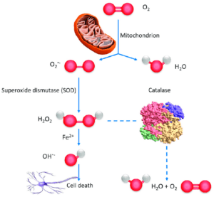
“Our body generates energy through various chemical reactions and during these processes, electrons are produced. These electrons are like tiny particles that carry energy. Inside our cells, there are powerhouses called mitochondria that receive these electrons and use them to create energy,” she explains.
However, sometimes, these electrons escape from their intended path and react prematurely with oxygen, creating unstable molecules called Reactive Oxygen Species (ROS). These ROS can be harmful to cells if they accumulate in large amounts.
ROS molecules can interfere with the normal communication networks in cells, known as signaling pathways. ROS molecules act as messengers within these pathways, relaying signals from one part to another. Some of these pathways control how cells grow and survive.
When there are changes in these pathways, it can lead to abnormal cell growth, which is a characteristic of cancer. ROS molecules play a role in these changes by affecting the signaling pathways.
Cancer vaccines: Can they pave the way towards a cancer-immune world?
QBET spectroscopy and how it works
A recent study at Harvard and UC Berkeley reported the advent of Quantum Biological Electron Transfer (QBET) spectroscopy, a new technique, that allows scientists to observe and study the movement of electrons in living cells in real-time.
This technique uses a special junction to create a pathway that enables scientists to see how electrons move through different molecules inside cells. By using this approach, the researchers came up with the idea of detecting electron transfer changes during cancer formation.
While the grant was applied for by Kannan, the project was ideated by Dr Rao.
The cancer researchers explored the application of QBET spectroscopy in detecting changes in electron transfer during the formation of cancer. They hypothesised that carcinogens can accept electrons and interfere with the normal pathways of electron transfer in cells.
“Expanding on the idea that cancer-causing substances can interrupt the flow of electrons in our cells, a new technique called QBET spectroscopic imaging has emerged. This method allows us to observe how electrons move around in living cells in a non-invasive and real-time manner. By doing so, it opens up exciting possibilities for improving cancer prognosis and diagnosis,” states Swetha.
Dr Vishal says that with QBET spectroscopic imaging, we can capture both the location and timing of electron movement within cells. This helps us understand the intricate quantum biological mechanisms involved in the transformation of healthy cells into cancerous ones. By studying these electron transfer dynamics, we can gain valuable insights into the early stages of cancer development. This technique holds promise for advancing cancer medicine by providing a way to detect and monitor cancer more effectively.
Their preliminary research on the same has been published in the journal Medical Hypothesis.
Also Read: Study says drinking hot tea may lead to oesophageal cancer
How will it help cancer patients?
By studying these changes in real-time using QBET spectroscopy, the researchers aim to develop a non-invasive method to detect and monitor cancer development.

This advancement could potentially lead to improved prognostic and diagnostic tools for cancer, providing a deeper understanding of the disease’s underlying mechanisms.
Researchers claim that this breakthrough in cancer detection opens up new possibilities for medical researchers and offers hope for improved outcomes for patients. Being able to observe and study electron transfer dynamics in living cells in real-time could revolutionise our ability to detect and understand the development of cancer.
With further advancements, QBET spectroscopy could become a powerful tool for early cancer detection, leading to more effective treatments and ultimately saving lives.
Current methods of testing to confirm the cancerous changes in tissues typically involve biopsies and biomarker tests. These are usually invasive in nature and only effective to detect a malignancy when established, as opposed to helping monitor pre-neoplastic/heoplastic changes.
Also, there are no accurate diagnostic tests in the Indian setting to help monitor the presence of neoplastic changes in patients with high stage cancers due to which prognosis of recurrent disease is very poor.
Also Read: AstraZeneca receives CDSCO approval for biliary tract cancer medicine
Collaboration with IISc
The Lee-Yung Family Fund, awarded to Swetha Kannan by Trinity Hall, has served incredibly useful in kickstarting pilot experiments of this project. Swetha says, “The prestige of the grant has further opened venues for collaboration on and expansion of the project’s experimental aims and deliverables.”
The researchers are currently working with IISc to develop and fine-tune their diagnostic tool and are hopeful of securing subsequent grants in the future to enhance its accessibility across India — particularly in rural areas where cancer diagnostics need to significantly improve in equitability and efficacy.
“Our long-term goal would indeed be to make QBET spectroscopy-mediated diagnostics accessible and affordable to the masses. We are currently optimising the tool for detection of head and neck cancers which are known to be caused by a certain set of carcinogenic compounds.”
However, the researchers are keen to explore the potential of the tool in detecting cancers that are associated with developmental/genetic defects (such as paediatric cancers) as opposed to those that arise from age-accumulated mutations and/or carcinogenic exposure.
Swetha adds, “We are incredibly passionate about and committed to making cancer care an equitable resource in India and our primary aim would be to make this diagnostic tool accessible to the (underprivileged) masses. It will certainly be a delight to have it find its space in the global diagnostic market, especially in other low and middle income countries.”