Infection
Zika study reveals how infection can cause microcephaly
Prenatal exposure to viruses capable of infecting the fetal brain, particularly in the first trimester, can cause a range of developmental defects in the baby. The Zika epidemic in Brazil during 2015–16 posed an extreme case, causing hundreds of babies to be born with microcephaly, or an abnormally small head.
Although cases have waned significantly, “the medical community agrees Zika could come back at any time, in any place where the mosquitoes that spread it live,” says Ganeshwaran Mochida, MD, MMSc, a specialist in microcephaly at Boston Children’s Hospital and a member of Boston Children’s Division of Genetics and Genomics.
And Zika is not the only virus of concern.
“Multiple viruses, including chickenpox, measles, cytomegalovirus, West Nile, and herpes simplex viruses can cause congenital brain diseases in newborns in the U.S.,” says neurobiologist Judith Steen, Ph.D., of the F.M. Kirby Neurobiology Center at Boston Children’s.
To try to get ahead of future outbreaks and prevent congenital developmental disorders caused by such neurotropic viruses, a team lead by Steen and Mochida set out to better understand how Zika affects the developing brain.
Combining proteomics (the study of proteins) and transcriptomics (the study of RNA), they identify potential targets for therapeutics that could protect babies’ developing brains. Their findings appear in the journal iScience.
An immune perspective
Most previous Zika research has overlooked one key factor in stunting brain growth: the immune system. In part, this is because mice, commonly used to model disease, aren’t very susceptible to the Zika virus, so researchers used mice with weakened immune systems that contract Zika more easily. Other studies have worked only with mouse nervous tissue grown from stem cells.
“To see the whole picture, you have to look at the whole organism in its natural state,” says Mochida, who helped track brain abnormalities during the outbreak in Brazil.
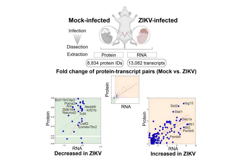
The team injected the Zika virus directly into the placentas of live pregnant mice with normal immune systems to mimic human Zika infection, which is transmitted from the mother’s blood through the placenta to the fetus. Through this method, the mouse embryos became infected early in their development.
The Steen laboratory then compared the RNA and proteins in the infected embryos’ brains to those in their healthy littermates. This revealed multiple molecular changes during the infection process that prevented normal brain development.
“No prior studies on Zika pathology have looked at both transcriptomics and proteomics with an immune perspective in an in vivo model,” Steen says.
Multiple barriers to brain development
The researchers found several things. First, Zika infection significantly up-regulated levels of immune proteins in the developing mouse brains, including MHC-1 proteins. This suggests that the virus triggers a substantial immune response that contributes to microcephaly.
“The immune system attacks cells infected with virus,” explains Steen. “If you can kill those cells, the outcome is usually good. But, if you start killing brain cells that are actively dividing brain cells, especially early in gestation, that can be devastating to brain development.”
Second, Zika infection lowered the levels of RNA and proteins related to the cell cycle—the process by which cells grow, replicate their DNA, and divide. By disrupting normal cell-cycle progression, Zika infection makes it impossible for the developing brain to grow properly.
The more viral particles and viral protein in the brain, the less of the normal cell-cycle proteins Steen and Mochida found.
Finally, Zika infection disrupted transcription factor signaling, altering the expression of genes involved in brain development. In Zika-infected brains, the team found abnormally low levels of many transcription factors that regulate the differentiation of neurons.
Therapeutic targets to avoid microcephaly?
Going a step further, the scientists looked to see which genes significantly up- or down-regulated by Zika could potentially be modulated with drugs to combat the virus’s effects.
The team identified 61 potentially targetable genes. Molecules targeting them could be tested in the mouse model at different stages to see if the disturbances the team observed are normalized, and whether any potential therapeutics would be safe for developing embryos.
Mochida hopes the team’s work will be important for understanding not just Zika, but how infection with neurotropic viruses affects the developing brain in general.
“We knew about the Zika virus for more than 50 years before the epidemic, but little research was done,” Mochida says. “We can learn from this experience how we will deal with the next epidemic. We shouldn’t forget.”
More information:
Kimino Fujimura et al, Integrative systems biology characterizes immune-mediated neurodevelopmental changes in murine Zika virus microcephaly, iScience (2023). DOI: 10.1016/j.isci.2023.106909
Children’s Hospital Boston
Citation:
Zika study reveals how infection can cause microcephaly (2023, August 9)
retrieved 9 August 2023
from https://medicalxpress.com/news/2023-08-zika-reveals-infection-microcephaly.html
This document is subject to copyright. Apart from any fair dealing for the purpose of private study or research, no
part may be reproduced without the written permission. The content is provided for information purposes only.

