
Cardiovascular
The evolving roles of TEE and ICE in structural heart interventions
Oct
Narang stressed that while ICE imaging is a powerful addition to the imaging toolkit, it is not likely to replace TEE entirely. Instead, the two technologies can work together effectively. ICE provides excellent 2D frame rates, but 3D imaging may have limitations in terms of frame rates and color quality. A collaborative approach between echocardiographers and interventionalists ensures comprehensive guidance during procedures.
He said there is a little bit of a learning curve using ICE as well, since the angles used for ICE are different from TEE. This requires a shift in the perspective of imagers. Communication between the imaging team and interventionalists is vital to achieve the best outcomes. The introduction of ICE catheters with digital steering capabilities is helping simplify this process, Narang explained.
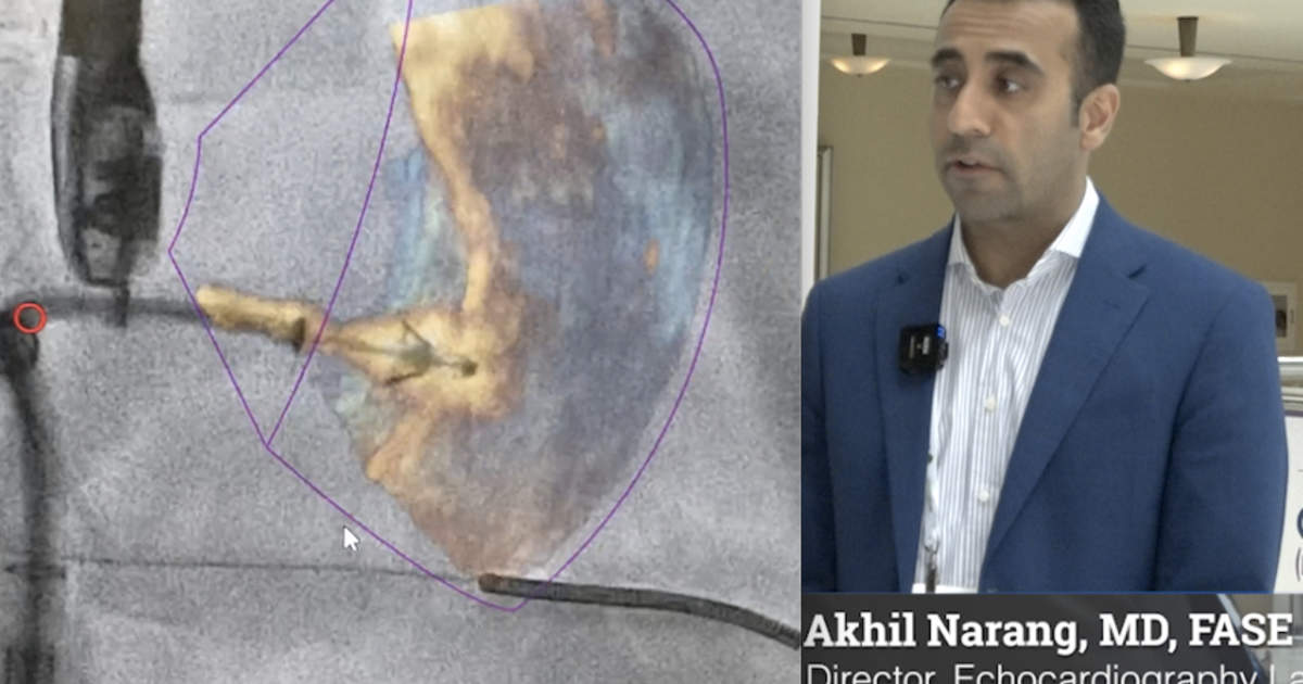
The posterior aspect of the tricuspid valve is where a lot of shadowing occurs from the TEE probe. Complex patients that have a mitral prosthesis, mechanical aortic valves also cause a lot of shadowing, Narang said.
“That posterior aspect is really difficult to get out from the shadows. And so ICE imaging directly in the right atrium can solve some of those problems. But, there are trade-offs for sure. So I think for the time being, we use both ICE and TEE in certain cases and I think it can be very helpful,” Narang explained.
The future of structural heart interventions
Northwestern has performed a large number of transcatheter tricuspid structural heart procedures because it has been involved in clinical trials for various devices. Narang highlighted the complexities of the tricuspid valve space, where patient selection and procedure planning require meticulous imaging. Tricuspid interventions often rely heavily on imaging due to the diverse anatomy of tricuspid valves. He said these lessons from trials centers will be important, because numerous structural heart experts predict devices for the tricuspid space will likely see FDA clearances in the next year or so, before any new mitral devices are cleared. Once this happens, there will likely be a rapid adoption of these new treatments for tricuspid valve regurgitation at structural heart centers.
Fusion imaging and co-registration to aid structural heart procedures
Fusion imaging, which combines echo and fluoroscopy or CT, plays a crucial role in visualizing paravalvular leaks and other structural heart issues, Narang said. As these technologies continue to develop, he said they will become increasingly important for guiding interventional procedures. Co-registration challenges are being addressed to make fusion imaging more efficient.
Real-time post-processing of images is also become more important. He said technologies like TrueView and GlassView for Phillips, and equivalent technologies from GE and Siemens, help show life-like surgical views of the anatomy using ultrasound, or can fade out anatomical structures so the underlying tissues can be seen. Both of these techniques help the operators see where they are at in the anatomy, what needs to be fixed and how to best navigate devices into positioning, he said.
“These allow us to really have a better understanding of the tissue characterization of the valves to really appreciate subtleties and flail segments or leaflets. I think the multi-planner reconstruction (MPR) technology has come a long way and a lot more simple, so that we’re able to do this pretty easily to get into an MPR view with high quality 2D reconstruction to guide procedures entirely,” Narang said.
One issue he sees with fusion technologies is that they work if you use one vendor’s echo system, co-registration system and their angiography system. But if you attempt to mix vendors the technology may not work. So every time you move, you have to reregister the image, he said.
He said automation of TEE functions is also getting better.
“I’m seeing a lot of companies that are investing time into doing measurements of the tricuspid valve and the Mitral valve. A lot is coming into that space to help guide the actual procedure and kind of make it more efficient so that is not a lot of time wasted when you’re trying to acquire optimal images, and then see them from multiple reconstructions,” Narang explained.
The expanding need for structural heart imagers
While echo automation, ICE and artificial intelligence being integrated into cardiac ultrasound systems may make it appear that there will be less of a need for interventional echocardiographers in the future, Narang said the opposite is true and demand will continue growing for this subspecialty.
Structural heart interventions, such as transcatheter aortic valve replacement (TAVR) and mitral valve repair with devices like the MitraClip, have rapidly gained acceptance over the past decade. TAVR now makes up more than 80% of aortic valve replacements in the U.S.
There are more mitral value replacement procedures performed each year than aortic, so when dedicated mitral transcatheter valves eventually become available there is an expectation they will follow a rapid adoption similar to TAVR. The tricuspid space is posed to expand rapidly in the next couple years. New types of structural heart interventions are also on the horizon.
“We see a huge demand. I think we’re still in the trial and early era of the tricuspid space, so there’s not many centers that are doing it. At Northwestern, we get a lot of referrals all throughout the country for these types of procedures, because we’re in numerous trials. I think when these devices become FDA approved, a lot of other places are going to do it. The tricuspid surgical outcomes historically have not been that great. They’ve had some higher challenges with morbidity and mortality. The preference might be to shift some of those patients that are considered higher risk to the transcatheter space,” he said.
Prioritizing structural heart imaging at ASE
Narang commended the ASE meeting for its dedication to highlighting structural heart interventions, making it a focal point for the imaging community. He discussed various sessions that emphasized the significance of these interventions and presented insights into the latest technologies and techniques.
Watch or read these two related interviews from ASE the current state of interventional imaging:
Interventional echocardiographers play a key role in the treatment of structural heart disease – Interview with Rebecca Hahn, MD
Interventional echocardiography expected to grow thanks to new structural heart procedures – Interview with Stephen Little, MD