
Congenital disorders
Ovotesticular disorder of sex development in a 46 XY adolescent: a rare case report with review of the literature
Oct
A 15-year-old patient, with no previous pathological history, raised as a boy, consulted the urology department of the University Hospital of Treichville for a right scrotal swelling, evolving for more than 5 years. The patient began full and normal male puberty at the age of 13.
The clinical examination revealed a patient in good general condition, measuring 1.56 m and weighing 47 kg, corresponding to a body mass index of 19.31 kg/m². The patient had Tanner stage 2 pubic hair, with a penis measuring 4 cm at rest, without hypospadias or cryptorchidism.
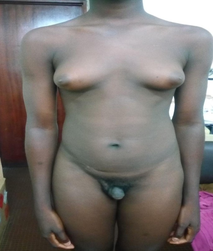
A painless, mobile and firm right scrotal mass was palpated. The left testicle was present but atrophic. He had a female morphotype, with marked gynecomastia (Fig. 1). The lymph nodes were free.
Male subject with bilateral gynecomastia
On pelvic and scrotal ultrasound, a right testicular solid tumor with a long axis of 6 cm was found, with absence of female genitalia. No uterus or adnexa was seen. The contralateral testis was without abnormality.
The biological workup consisted of TESTOSTERONE (0.1 ng/ml), ESTRADIOL (20 ng/ml), ALPHA-FETO-PROTEIN (5 ng/ml) and β HCG (< 01ui /l); all of which came back normal.
A right orchiectomy was performed and the specimen sent to the pathology laboratory.
Macroscopically, it was a lumpy mass. On section, there were several whitish nodules surrounded by a yellowish border (Fig. 2).

Whitish multinodular areas and whitish territories surrounded by a yellowish border
The resected specimen was fixed with 10% formaldehyde, followed by conventional dehydration, kerosene embedding, sectioning and hematoxylin and eosin (HE) staining. On histology, there was a juxtaposition of ovarian stroma with ovarian follicle and seminiferous tubules (Figs. 3 and 4, and 5). In view of this aspect we retained the diagnosis of ovotestis. The karyotype was determined to be male (46, XY), confirming the diagnosis of ovotesticular disorders of sex development. An initial interview was held with the parents in the presence of a psychologist to explain their child’s pathology, before the child was informed during a second interview in the presence of the parents and the psychologist.

Juxtaposition of ovarian stroma and seminiferous tubules (HESx100)

Ovarian tissue with a follicular cyst and an ovarian follicle (HESX100)

Testicular parenchyma seen at high magnification (HESX100)
Hormonal therapy based on 80 mg testosterone undecanoate capsules at a dose of once a day and mastectomy were proposed, but the patient was lost to follow-up.