Infection
Q&A: New imaging method enables quick lung infection identification
November 02, 2023
5 min read
Key takeaways:
- Chest X-rays are not a quick way to diagnose and treat patients with suspected lung infections.
- An imaging technique is currently being developed to rapidly identify lung infections.
With the current chest imaging technology, patients with suspected lung infections are unable to receive a specific diagnosis unless they undergo an invasive procedure and wait 2 to 3 days for their results, according to a press release.
However, Nalinikanth Kotagiri, PhD, MBBS, associate professor of pharmaceutical sciences at University of Cincinnati James L. Winkle College of Pharmacy, is hoping to change this by assessing various kinds of injectable probes used during chest imaging and creating a new imaging method with his findings.
This research, supported by a grant from the National Heart, Lung, and Blood Institute, will help expediate the process of diagnosing infections and lead to earlier treatment.
Healio spoke with Kotagiri to learn more about his research, how the proposed new imaging technique will benefit both patients and clinicians and how it can continue to be used after treatment.
Healio: What inspired your idea to research the effectiveness of different injectable probes used during chest imaging?
Kotagiri: COPD is a chronic illness that can have periodic flare-ups, characterized by acute worsening of symptoms. These flare-ups accelerate the progressive decline in lung function in patients with COPD, deteriorating health-related quality of life and, when associated with ventilatory failure, to premature death. Depending on region and country, the bacteria frequently encountered as a cause of flare-ups is Pseudomonas aeruginosa. P. aeruginosa infections also appear to portend a worse prognosis with higher 30‐ and 90‐day mortality rates compared with flare-ups not associated with this bacterium. Therefore, in such patients, an accurate and timely diagnosis is required to identify, or exclude, bacterial pathogens, which may present initially or arise later in the course of the disease.
Healio: Although chest X-rays can be a useful tool to diagnose patients, what are their limitations?
Kotagiri: Conventional X-ray imaging is common in such instances; however, there are many challenges.
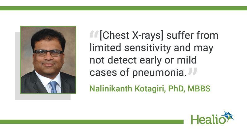
They suffer from limited sensitivity and may not detect early or mild cases of pneumonia, especially if the infection is located in the central parts of the lung. The resolution is not as high to pick finer details, making interpretation of the images rather difficult. If there are other co-existing conditions associated with the heart or lungs it can confuse the radiographic picture. In instances where the flare-ups are caused by viral infections, they may present a picture similar to bacterial infections, making treatment decisions challenging. Follow-up microbiological or molecular testing is still required after performing imaging to establish a diagnosis, resulting in a prolonged, uncertain process that takes several days.
Healio: How will this proposed new imaging method benefit both patients and clinicians? What lung/respiratory conditions could be identified with this method?
Kotagiri: Nuclear imaging, particularly positron emission tomography (PET), is a noninvasive imaging modality that is routinely used to diagnose lesions with high sensitivity and superior contrast. Therefore, this modality allows for exciting possibilities in imaging bacteria and potentially permitting their accurate detection and quantitation. However, imaging of “live” bacteria in the body during infection remains a continuing challenge. Imaging of live bacteria would provide information regarding presence, location, quantity, survival and status of pathogenic bacteria, such as P. aeruginosa in animals or patients with high sensitivity and contrast at any desired time-point following administration of the contrast agent.
We anticipate that our technique will be able to: (1) identify subclinical bacterial colonization in patients with acute flare-ups, to determine if the bacteria that are ultimately responsible for the infection are the same strains that was found to be colonizing, particularly for P. aeruginosa due to the high mortality associated with this bacteria; (2) distinguish flare-ups of bacterial origin than those caused by viruses; and (3) facilitate antibiotic treatment monitoring in patients. Ideally, such an approach should also be able to determine source and distribution of the pathogens throughout the lung and potential spread to other parts of the body, to minimize indiscriminate use of broad-spectrum antibiotics and adopt a more focused treatment strategy targeting specific pathogens.
Healio: How are you studying this new method and what have you found so far?
Kotagiri: Transition metal ions are essential micronutrients for all life forms. Bacteria have developed sophisticated mechanisms for metal acquisition and transport to maintain metal balance inside cells. They do this through metal-binding molecules known as siderophores and dedicated transporters on their cell membrane that selectively bind to these siderophores that have evolved to precisely regulate this process. Siderophores exhibit high affinity and selectivity to the bacteria with each type of bacteria manufacturing siderophores unique to them. This selectivity in siderophore uptake into their unique bacterial strains is what we exploit to image bacteria such as Pseudomonas, including other species such as Escherichia coli and Klebsiella. The contrast agent is prepared by complexing or mixing the siderophores with a radiometal, such as copper-64. JCI Insight recently published our proof-of-concept findings in animal infection models that shows high specificity of the siderophore, Yersiniabactin to E. coli and Klebsiella, and have recently identified unique siderophores specific to Pseudomonas species.
The grant will allow us to explore further development and optimization of this technology in animal models of COPD with an overlaying bacterial infection as well as co-infection models that have both bacterial and viral infections.
Healio: In the press release, you noted that this imaging method could also be used following treatment. How will further imaging help patients with lung diseases improve?
Kotagiri: Current technologies fail to provide rapid feedback about the effect, or adequacy, of a selected antimicrobial regimen. Because this technology allows us to image “live” bacteria, as dead bacteria do not show up in the scans, we would be able to know whether the bacteria causing the infection is actively proliferating in the lungs and elsewhere in the body. In the event the prescribed antibiotic regimen is not effective in eliminating the bacteria, the scan should be able to reveal not only the location and presence of the surviving bacteria but also quantitate the bacterial burden because the scans are quantitative. This could potentially help the clinicians in deciding whether to either change the antibiotics or increase the dose. This could be particularly useful in cases where they are dealing with multidrug resistant bacteria, where timely decision making to switch to last-resort antibiotics could prove to be critical for treatment outcomes.
Healio: Why is it important to reevaluate and improve upon existing imaging techniques?
Kotagiri: While there are other PET contrast agents, such as Fluorine-18 labeled glucose (18FDG) that is considered a gold-standard in the clinics, recent studies have shown that PET imaging with these radiopharmaceuticals enables detection of infection not because they image live bacteria but because they label inflammatory leukocytes or white blood cells that infiltrate the infection site. 18FDG-PET, however, does not allow for the discrimination between infective and inflammatory processes (such as cancer) and does not have the ability to distinguish one genus/species/strain of bacteria from another. Moreover, 18FDG-PET is dependent on host inflammatory responses to infection, which may be reduced or absent in immunosuppressed patients who are most at risk for infection. Our technique that labels live bacteria directly circumvents these issues and could potentially provide an alternative modality for imaging infections in the clinic as well as an important biomedical tool to assist investigators doing basic and translational research to better understand host-microbiome/bacterial interactions.

