Cardiovascular
The expanding scope of FDG-PET in nuclear cardiac imaging
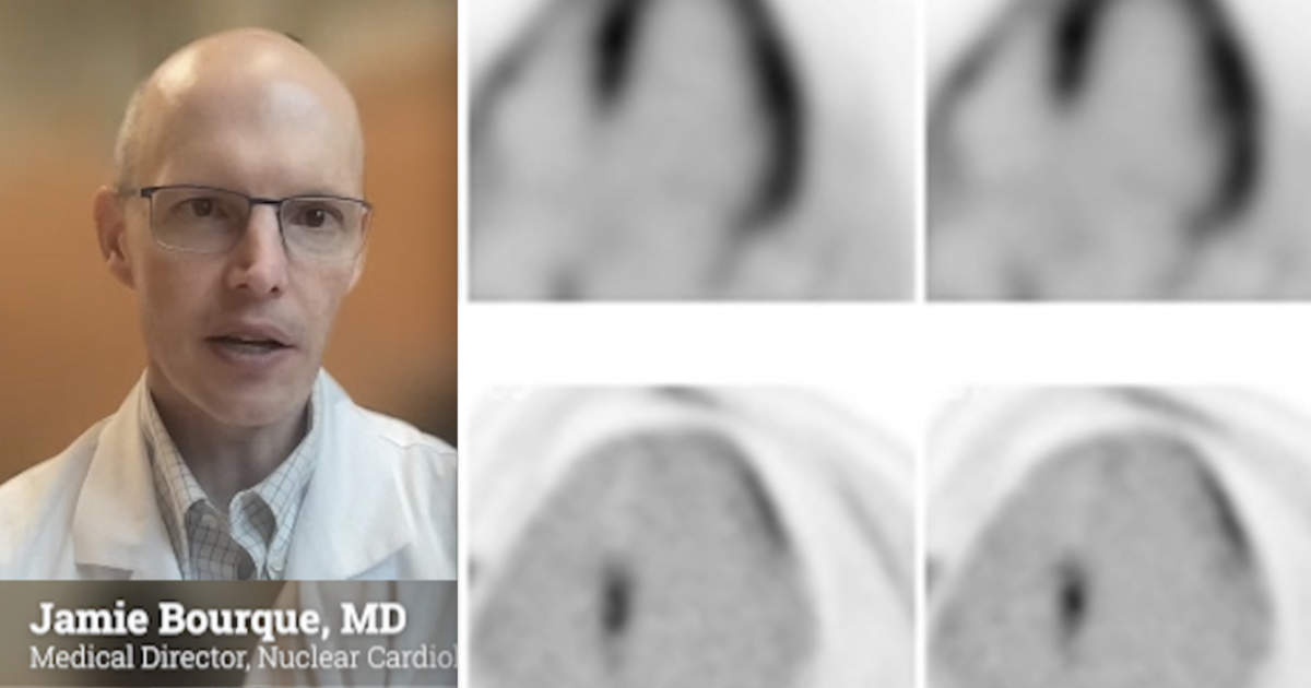
FDG-PET imaging is typically associated with the detection of metabolically active malignant lesions in various cancers, but the American Society off Nuclear Cardiology (ASNC) 2023 meeting shed light on the expanding horizons of FDG-PET in cardiovascular imaging. Cardiovascular Business spoke with Jamie Bourque, MD, medical director of the nuclear cardiology and stress laboratory and medical director of the echocardiography lab at the University of Virginia, who spoke at ASNC on FDG imaging.
“We are excited about increasing applications in cardiovascular infection, in vasculitis, for refined use in myocardial viability, to assess for inflammation as in disease states such as cardiac sarcoidosis and vasculitis to ability to diagnose and assess cardiovascular infection. It’s a ‘jack of all trades’ and [this is] an exciting time for cardiac PET,” Bourque explained.
FDG-PET’s increased availability owes much to the rapid expansion of cardiac PET cameras. Now, these cameras serve dual roles, enabling stress testing and facilitating the use of FDG in new cardiac imaging roles.
FDG enables inflammation and infection imaging
In clinical practice, FDG-PET plays a pivotal role in assessing inflammatory conditions, identifying cardiac involvement in sarcoidosis, evaluating disease progression, and gauging therapeutic responses. Additionally, it aids in diagnosing cardiovascular infections, providing insights into endocarditis, embolic phenomena and complications related to cardiac electrophysiology (EP) devices.
“It is particularly helpful when someone has possible endocarditis by existing clinical criteria and other imaging modalities. The mainstay for imaging of endocarditis continues to be echocardiography, but it is not always a perfect technique. And so occasionally FDG PET-CT can be used to make a diagnosis of endocarditis. It also can help identify whether there are complications outside of the actual valve itself in valvular endocarditis, such as someone who may have a periprosthetic abscess for instance,” Bourque explained.
For EP device infections, it can be very helpful not only for identifying if there is infection of a cardiac lead and specifically which areas of the device are infected, but particularly for assessment of pocket infections. If there is more superficial inflammation, this would not necessarily require a device to be taken out, versus someone who has a deep pocket infection with presumed involvement of the device itself. In that case he said the device would typically need to be removed. This could be critical for patients who are device dependent or if they are frail, because removal of the device and leads may cause substantial complications, or it may be challenging to remove, Bourque said.
One of FDG-PET’s distinguishing features lies in its glucose analog nature. It mirrors glucose uptake patterns, enabling myocardial viability assessment and identifying inflammation and infection by targeting metabolically active cells. This versatility allows for a comprehensive assessment of various cardiovascular conditions. Since white blood cells exclusively use glucose for energy, FDG can help show areas of infection.
FDG agents have longer half-lives than rubidium for cardiac imaging
The ease of acquiring FDG radiotracers commercially is enabled by their relatively long half-life of 110 minutes. This enables wider spread use because it can be made at a commercial cyclotron up to a couple hours away and transported to the imaging center.
“The other advantage to that is FDG is used extensively in oncology, so most institutions will have some sort of sourcing already available for FDG for their oncologic purposes. And so it’s easier for cardiology to tack on to that existing framework,” Bourque said.

