
Congenital disorders
OCT-based approach examines human physiology
Nov
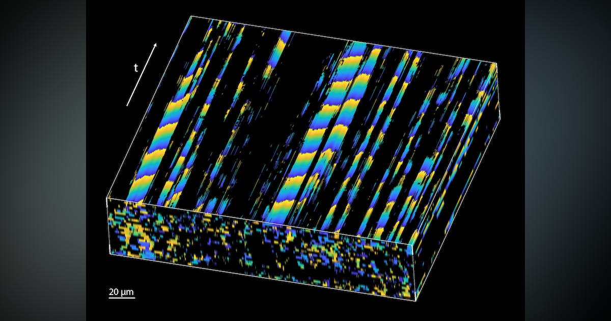
A metachronal wave—wavy movements produced by the sequential action of structures like cilia—is created by phase delays in periodic beating between neighboring cilia. This type of phase mapping allows the researchers to visualize and quantify the metachronal wave created by cilia.
Why OCT?
For their study, Larina says OCT was their top-choice starting point because it can best facilitate the study of biological processes in vivo.
“It allows for dynamic volumetric visualization of biological samples with a resolution of a few micrometers (less than the size of one cell) at a depth of 1 to 3 millimeters,” Larina says. “Since it relies on natural tissue contrast and does not require labels, OCT is perfect for in vivo applications.”
Conventionally, high-speed video microscopy is used to expose ciliated surfaces and analyze their dynamics. It’s a more invasive technique that typically involves dissecting an organ of interest and exposing the ciliated surface for imaging. This disrupts the physiological environment of the cilia, resulting in an inaccurate interpretation.
OCT has been part of multiple approaches for motile cilia research and is used for cilia location mapping, cilia beat frequency analysis, and mapping microflows induced by cilia. But the team’s new approach is the first that enables analysis of cilia coordination (metachronal wave) through tissue layers.
“This is important because cilia are working in coordination to move cells and mucus, and proper coordination—not just their beat frequency—is required for the transporting function,” Larina says. “There is currently no other technology that can reach such depth-resolved measurements.”
The research
Ciliary motility is used by microorganisms as well as more complex species including humans; it’s critical for survival. “A complete disruption of ciliary dynamics leads to very early embryonic lethality,” Larina explains. “While subtle abnormalities in ciliary dynamics (motile ciliopathies) lead to serious clinical complications affecting different organ systems.”
Investigation of motile cilia function and regulation “is important for the advancement of healthcare,” Larina says.
Imaging ciliary dynamics is challenging, however, due to motile cilia’s small size. Investigating ciliary beat coordination without directly exposing the ciliated surface was not technologically possible until now.
“Our innovation allows us to map cilia metachronal wave propagation within the lumen of a mouse fallopian tube without disrupting the normal physiological environment, which was not previously accessible,” Larina says, and points out that researchers can now map and quantify at micro-scale spatial resolution with a depth-resolved field-of-view (see Fig. 3).