Congenital disorders
Erythema multiforme major associated with SARS-CoV-2 infection in a patient with skin of colour
A 38-year-old man with Fitzpatrick type V skin presented to the emergency department with a 2-week history of fever, productive cough, rhinorrhea and 5 days of oral pain. He was otherwise healthy and took no medications. He had been vaccinated against SARS-CoV-2, but SARS-CoV-2 infection was suspected and confirmed with a nasopharyngeal swab.
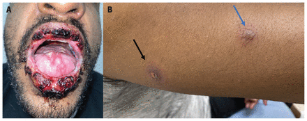
Examination revealed erosive plaques to the oral mucosa and glans penis, as well as hemorrhagic crusting of the lips (Figure 1A). Lesions on the upper and lower extremities had targetoid plaques with 3 concentric zones — a central necrotic region, a palpable edematous halo and a well-circumscribed erythematous ring — typical lesions of erythema multiforme major ( Figure 1B). However, atypical erythematous– violaceous lesions featuring 2 concentric zones with central bullae and indistinct outer borders made the clinical diagnosis unclear. Therefore, we performed punch biopsies and direct immunofluorescence of adjacent nonlesional skin to rule out autoimmune blistering conditions. Histology showed subepidermal blistering with necrotic keratinocytes, consistent with erythema multiforme major secondary to SARS-CoV-2 infection. Our patient’s systemic and cutaneous symptoms resolved with a 3-week tapering regimen of oral prednisone (starting with 40 mg/d), mometasone furoate (0.1% cream) to the lips, benzydamine and diphenhydramine hydrochloride– dexamethasone–nystatin mouthwashes (3 times daily).
” data-icon-position data-hide-link-title=”0″>
A 38-year-old man with SARS-CoV-2–associated erythema multiforme major. (A) Mucosal examination revealed erosive plaques to the ventral tongue, accompanied by erosions on the buccal mucosa and lips with overlying hemorrhagic crusting. (B) The patient’s right posterior arm had a classic targetoid plaque with 3 concentric zones, namely a de-roofed vesicle adjacent to an edematous blanchable ring and a well-circumscribed red ring on the periphery (black arrow). The blue arrow illustrates an atypical targetoid plaque with 2 concentric zones of a central bulla and poorly defined outer borders.
Erythema multiforme major is an immune-mediated, self-limited type IV hypersensitivity condition, characterized by mucosal erosions and targetoid plaques.1 The differential diagnosis includes fixed drug eruptions, Stevens–Johnson syndrome, autoimmune blistering disorders (e.g., mucous membrane pemphigoides, paraneoplastic pemphigus) and reactive infectious mucocutaneous eruption. Currently, no guidelines exist for treating SARS-CoV-2–associated erythema multiforme major. For erythema multiforme major with severe mucosal involvement, consensus supports topical and systemic corticosteroids, and antiseptic mouthwashes to manage potential sequelae, including dysphagia, dehydration and hospital admission.1,2
As highlighted in this report and a recent review article, patients with skin of colour can present with mucocutaneous manifestations that differ in appearance compared with lighter skin.3 Although publications on SARS-CoV-2–associated cutaneous manifestations are increasing, patients with skin of colour remain underrepresented, potentially delaying diagnosis and treatment.3
Footnotes
-
Competing interests: None declared.
-
This article has been peer reviewed.
-
The authors have obtained patient consent.
This is an Open Access article distributed in accordance with the terms of the Creative Commons Attribution (CC BY-NC-ND 4.0) licence, which permits use, distribution and reproduction in any medium, provided that the original publication is properly cited, the use is noncommercial (i.e., research or educational use), and no modifications or adaptations are made. See: https://creativecommons.org/licenses/by-nc-nd/4.0/

