
Skin Health
Actinic Keratosis
Dec
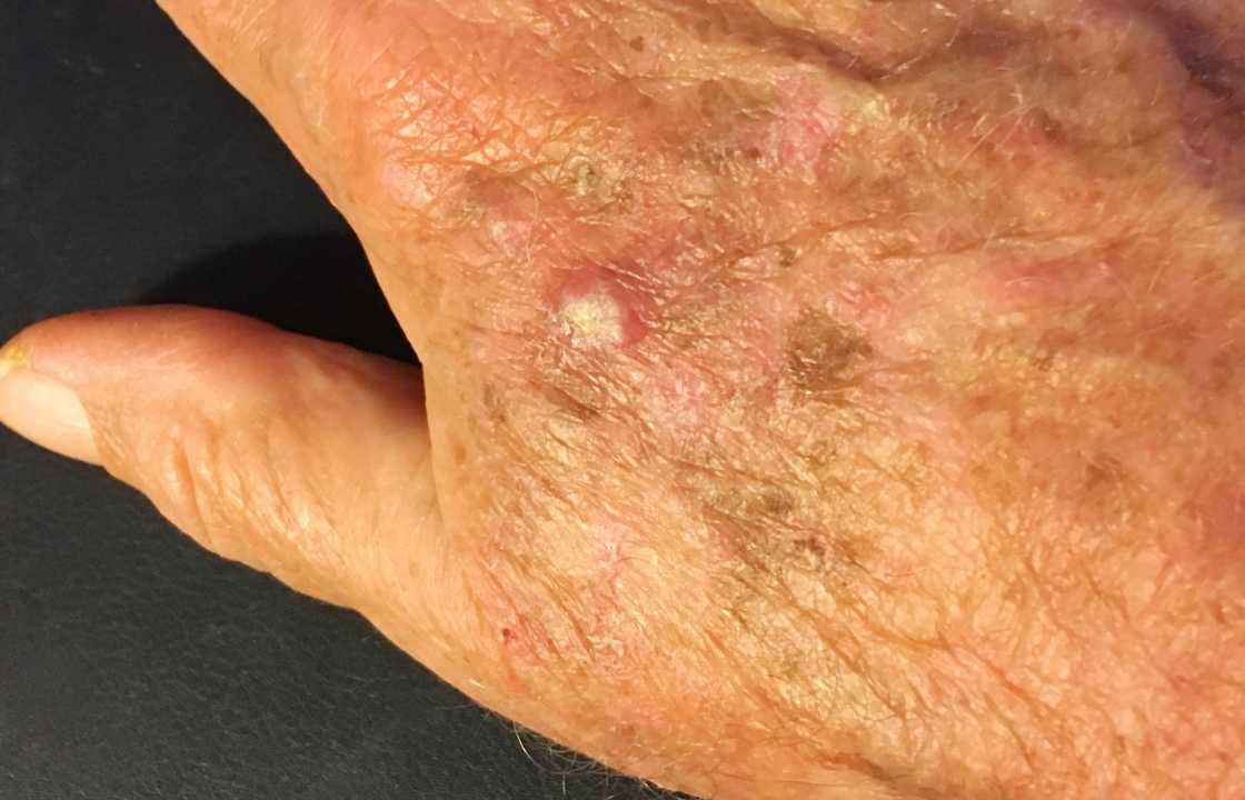
An actinic keratosis (ak-TIN-ik ker-uh-TOE-sis) emerges as a testament to the intricate relationship between prolonged sun exposure and the skin’s response to environmental stimuli. This condition manifests as rough, scaly patches on the skin, presenting a visual narrative of years spent under the sun’s embrace. Frequently encountered on the face, lips, ears, forearms, scalp, neck, or back of the hands, actinic keratosis serves as a reminder of the profound impact of ultraviolet (UV) rays on our skin’s health.
Decoding Actinic Keratosis
Actinic keratoses, colloquially known as solar keratoses, embody scaly spots or patches residing on the skin’s topmost layer. With the passage of time, these patches may undergo a transformation, developing a hardened, wartlike surface that adds an extra layer of complexity to their visual manifestation.
Commonly making their debut in individuals over the age of 40, actinic keratoses unfold their narrative gradually, emphasizing the importance of minimizing sun exposure and adopting protective measures against UV rays. Left unchecked, the risk associated with actinic keratoses taking a potentially ominous turn towards squamous cell carcinoma, a form of skin cancer, looms between 5% to 10%.
Symptomatic Portrait
Actinic keratoses exhibit a spectrum of appearances, with symptoms encapsulating:
1. Rough, Dry, or Scaly Patches: Typically measuring less than 1 inch (2.5 centimeters) in diameter, these patches manifest as a tactile testament to the skin’s response to prolonged sun exposure.
2. Flat to Slightly Raised: The patches or bumps on the skin’s surface can range from flat to slightly raised, creating a diverse landscape of textural variations.
3. Wartlike Surface: In some instances, actinic keratoses evolve to feature a hard, wartlike surface, adding an extra layer of complexity to their presentation.
4. Color Variations: The color palette of actinic keratoses includes shades of pink, red, or brown, contributing to their visual diversity.
5. Sensory Signals: Itching, burning, bleeding, or crusting may accompany these patches, creating a sensory interplay that adds nuance to the symptomatic journey.
6. Localization: New patches or bumps tend to emerge on sun-exposed areas of the head, neck, hands, and forearms, aligning with their etiological connection to UV radiation.
Navigating the Evaluation Terrain
Given the challenge of distinguishing between noncancerous spots and their potentially cancerous counterparts, seeking professional evaluation becomes paramount. Healthcare providers are adept at discerning subtle changes, especially if a scaly spot or patch persists, grows, or exhibits signs of bleeding.
Roots of Actinic Keratosis
The genesis of actinic keratosis lies in the frequent or intense exposure to UV rays from the sun or tanning beds. While its occurrence is not confined to a specific demographic, certain risk factors elevate susceptibility:
1. Physical Characteristics: Individuals with red or blond hair and blue or light-colored eyes are at an increased risk.
2. Sun Exposure History: A history of extensive sun exposure or sunburn amplifies the likelihood of actinic keratosis development.
3. Skin Response: Those prone to freckling or burning upon sunlight exposure face an elevated risk.
4. Age: The risk escalates with age, with individuals over 40 more prone to its manifestation.
5. Geographic Location: Residing in sun-drenched locales or working outdoors accentuates the risk.
6. Immune System Health: Individuals with a weakened immune system face an augmented risk profile.
Potential Complications
Early intervention is pivotal in mitigating the impact of actinic keratosis. If left untreated, however, a subset of these patches might progress to squamous cell carcinoma. Detecting and treating these early stages of skin cancer is generally not life-threatening, underscoring the importance of proactive management.
Sun Safety and Preventive Measures
Embracing sun safety measures serves as a cornerstone in preventing actinic keratosis. The following steps contribute to shielding the skin from the deleterious effects of UV radiation:
1. Time Limitation: Limiting sun exposure, especially during the peak hours between 10 a.m. and 2 p.m., and avoiding prolonged exposure leading to sunburns or suntans.
2. Sunscreen Application: Regular application of a broad-spectrum, water-resistant sunscreen with an SPF of at least 30, as recommended by the American Academy of Dermatology. This extends to all exposed skin, including the lips, with reapplication every two hours or more frequently during water activities or excessive sweating.
3. Protective Attire: Donning tightly woven clothing that covers the arms and legs, coupled with a broad-brimmed hat for enhanced sun protection. This surpasses the efficacy of baseball caps or golf visors.
4. Tanning Bed Avoidance: Steering clear of tanning beds, recognizing the comparable skin damage induced by UV exposure in both natural and artificial settings.
5. Regular Skin Checks: Vigilant self-examination of the skin, reporting any changes to healthcare providers promptly. This includes regular checks of the face, neck, ears, scalp, arms, and hands.
Diagnostic Expedition
Diagnosing actinic keratosis often involves a visual examination by healthcare providers. While the characteristic presentation may suffice for a conclusive diagnosis, additional tests, such as a skin biopsy, may be pursued if uncertainties persist. A skin biopsy entails the extraction of a small skin sample for laboratory analysis, often performed in a clinic setting after administering a numbing injection.
Post-treatment, regular skin checks, at least once a year, are typically recommended to monitor for potential signs of skin cancer.
Treatment Modalities
Actinic keratosis exhibits a dynamic response to various treatment modalities, with the choice dependent on factors such as the extent of involvement and individual characteristics. Common approaches include:
1. Topical Medications: Health care providers may prescribe medicated creams or gels, such as fluorouracil, imiquimod, or diclofenac, for the removal of multiple actinic keratoses. These products may induce temporary skin inflammation, scaling, or a burning sensation.
2. Cryotherapy: A prevalent method involving the freezing of actinic keratoses with liquid nitrogen, leading to blistering or peeling. The healing process results in the shedding of damaged cells, allowing new skin to emerge. Potential side effects include blisters, scarring, changes in skin texture, infection, and alterations in skin color.
3. Curettage and Electrosurgery: The use of a curet to scrape off damaged cells, followed by electrosurgery using an electric current to cut and destroy affected tissue. This procedure necessitates local anesthesia, with potential side effects encompassing infection, scarring, and changes in skin color.
4. Laser Therapy: An evolving technique employing ablative laser devices to obliterate actinic keratosis, facilitating the emergence of new skin. Side effects may include scarring and discoloration of the affected skin.
5. Photodynamic Therapy: Involving the application of a light-sensitive chemical solution to the affected skin, followed by exposure to a specialized light source to destroy actinic keratosis. Side effects may include inflamed skin, swelling, and a burning sensation during therapy.
Preparing for the Healthcare Odyssey
Embarking on the journey of seeking medical attention for actinic keratosis involves an initial interaction with primary care providers. In certain scenarios, direct referral to dermatologists, specialists in skin diseases, might be recommended.
Patient Preparations
To optimize the healthcare encounter, preparing a list of questions becomes a valuable strategy. For individuals grappling with actinic keratosis, key queries include:
1. Diagnostic Confirmation: Inquiring about the necessity of additional tests to confirm the diagnosis.
2. Treatment Options: Exploring the diverse treatment options available, along with a comprehensive understanding of the pros and cons associated with each.
3. Financial Considerations: Delving into the financial aspects of treatments, including the costs involved and potential coverage by medical insurance.
4. Vigilance Guidelines: Seeking guidance on the signs of suspicious changes in the skin post-treatment, facilitating proactive monitoring.
5. Follow-up Expectations: Understanding the anticipated follow-up procedures and their frequency.
Physician Interactions
On the physician’s end, the exploration into the patient’s history involves a series of questions aimed at unraveling the nuances of the condition:
1. Onset Details: Inquiring about when the patches or spots were initially noticed.
2. Multiplicity: Assessing whether multiple patches or spots have manifested.
3. Evolutionary Changes: Exploring any alterations observed in the affected skin over time.
4. Impact on Well-being: Gauging the extent to which the condition is bothersome to the individual.
5. Sun Exposure History: Delving into the frequency of sunburns or severe sunburn episodes.
6. UV Radiation Exposure: Evaluating the routine exposure to sun or ultraviolet (UV) radiation, along with protective measures adopted.
Conclusion
In the expansive realm of actinic keratosis, understanding its nuances empowers individuals to navigate the complexities of prevention, diagnosis, and treatment. As the sun remains a constant presence in our lives, embracing sun safety practices emerges as a formidable shield against the evolution of actinic keratosis into more ominous stages. With the collaborative efforts of healthcare providers and vigilant individuals, the narrative of actinic keratosis transforms from a potential precursor to skin cancer to a tale of proactive management and skin well-being. May this guide serve as a beacon, illuminating the path towards sun-conscious living and skin health.