Infection
Smartphone-based imaging reveals a breakthrough in identifying infected wounds
A recent Frontiers in Medicine study assesses the effectiveness of hyperspectral imaging (HSI) to identify differences between infected and non-infected wounds.
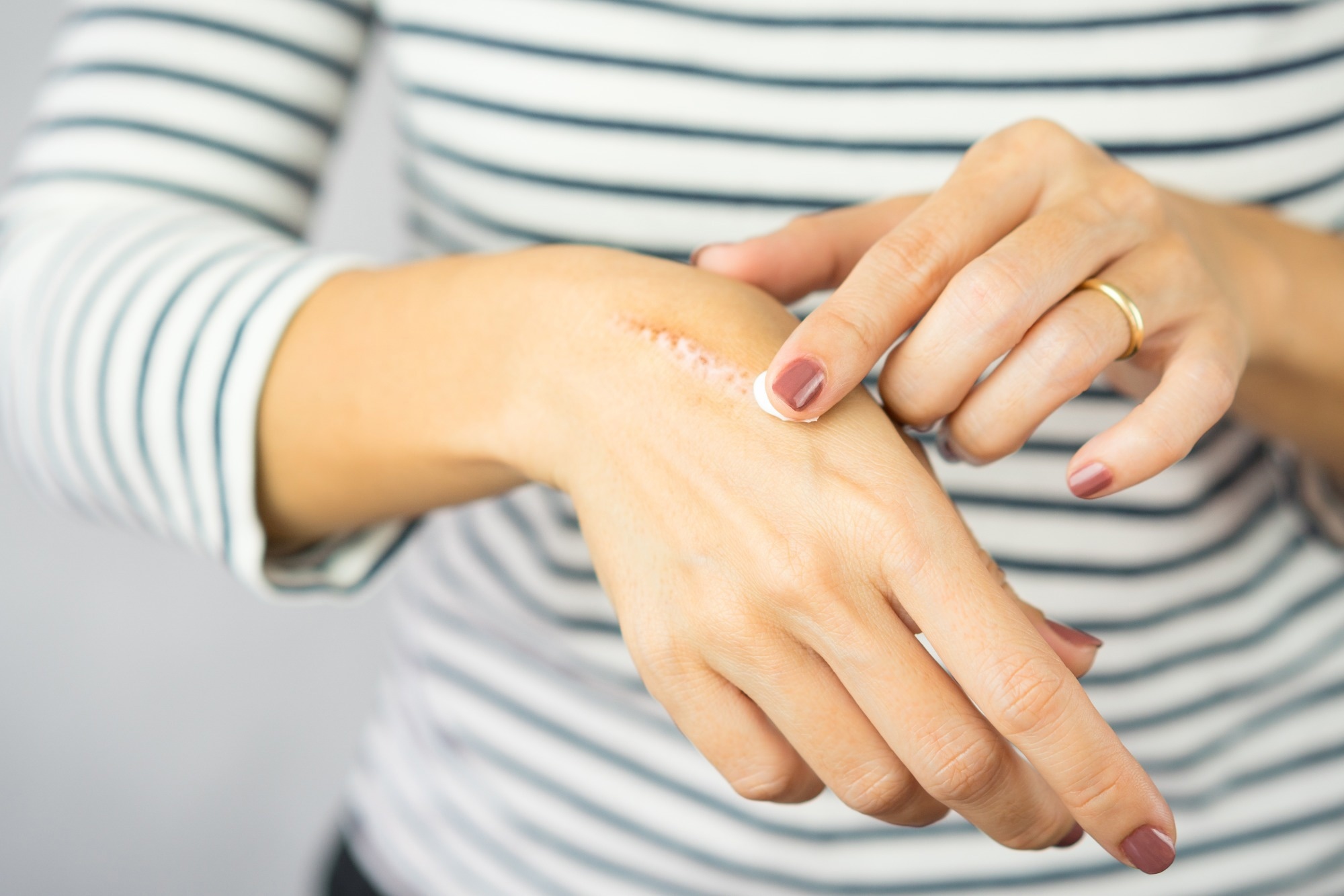
Study: Is my wound infected? A study on the use of hyperspectral imaging to assess wound infection. Image Credit: myboys.me / Shutterstock.com
Background
The three main components of wound healing are precursor cells that can multiply and differentiate into fibroblasts and keratinocytes, neo-angiogenesis to reinstate normal blood flow to the injured site, and the immune system to control inflammatory reactions. IWound healing is compromised or delayed if one of these components functions less efficiently,
Infections commonly induce the development of chronic wounds that decelerate the healing process. In chronic wounds, the healing process does not progress beyond the inflammatory phase of wound healing.
If the skin or wound is in non-sterile environments, a wide-ranging infection could occur, including colonization, local infection, contamination, and systemic infection. Thus, it is imperative to identify the type of infection to provide proper treatment. However, the accuracy of identifying the specific type of wounds based on clinical inspection is below 60%; therefore, there is an urgent need for more accurate diagnostics to detect infected wounds.
Although more accurate molecular and culture techniques are available, they are often expensive, time-consuming, and inaccessible to all. Thus, microbiological assessment methods are often used to determine infection.
Recently, infrared thermography (IRT) has shown great promise in diagnosing inflammation and infection in wounds. Although this method cannot detect the presence of an infectious process, the incorporation of violet light has facilitated bacterial fluorescence (BF) in wounds.
The BF method can only identify bacterial presence on the wound surface, as this imaging technique penetrates less than 1.5 millimeters (mm) into the tissue. Therefore, this diagnostic method is prone to miss any bacterial contamination beyond the aforementioned depth. Furthermore, it cannot detect infection due to fungal agents.
The Swift Ray 1 is a novel hyperspectral imaging (HSI) device that enables the acquisition of images through a smartphone for medical diagnosis. HSI provides diagnostic information regarding the morphology, composition, and physiology of the tissue by acquiring a hypercube that is a multi-dimensional image dataset.
The Ray 1 device contains violet light sources, near- and long-wave infrared sensors, and visible range LEDs that allow for the acquisition of visible light, IRT, and BF images as a hypercube. This device is also equipped with the Swift Skin and Wound app that enables accurate wound area measurement, fluorescence area quantification, and temperature quantification.
About the study
The current study hypothesizes that wounds can be categorized by analyzing the HSI data acquired by the Ray 1 device. These wound categories could be associated with an inflammatory responses, not asociated with an inflammatory response, or being infected. The current study aimed to determine whether HSI images could be used to differentiate between infected and non-infected wounds.
The study cohort consisted of patients with acute or chronic wounds. All relevant wound images were acquired by trained surgeons using a smartphone containing a Ray 1 HSI camera along with a Swift Skin and Wound app. The sample contained clinical photographs (visible light images), BF images, and thermograms.
Based on the International Wound Infection Institute Wound Infection Continuum (IWII-WIC) recommendations, a wound is considered infected by clinicians in the presence of local infection. Typically, infected wounds are treated with antibiotics.
Before imaging, the wound was cleaned with saline solution and patted dry. If required, loose skins and blisters were removed and the wounds were allowed to reach room temperature for five minutes.
While acquiring HSI and BF images, the camera was placed at 15 cm and a 90° angle away from the wound bed. Thermal imaging was performed following standard protocol.
Study findings
A total of 66 patients were recruited in this study and 20 wounds were identified as clinically infected. A combination of patient clinical data, IRT, visible light, and BF imaging was able to differentiate between infected and non-infected wounds.
The current study also analyzed temperature gradient patterns to assess the body’s response to the wound. Diagnosis of the type of infection based on only temperature analysis could be challenging.
The current method enabled the prediction of infected, inflamed, and non-infected wounds with 74% accuracy. As compared to all types, this approach performed best on infected wounds, with 100% sensitivity and 91% specificity. Comparatively, this method detected non-inflamed wounds with 94% sensitivity and 70% specificity, whereas inflamed wounds were associated with 85% sensitivity and 77% specificity.
Conclusions
The combinational approach involving a pocket-size HSI imaging device enabled point-of-care assessment, as well as the differentiation of inflamed, infected, or non-infected wound types, with increased sensitivity and specificity of infection detection. Precise diagnostic information will help clinicians to provide more targeted treatment.
- L., J., A., M., Fraser, R. D., Bartlett, R., et al. (2023). Is my wound infected? A study on the use of hyperspectral imaging to assess wound infection. Frontiers in Medicine 10. doi:10.3389/fmed.2023.1165281

