
Cancer, Health Care, Insight
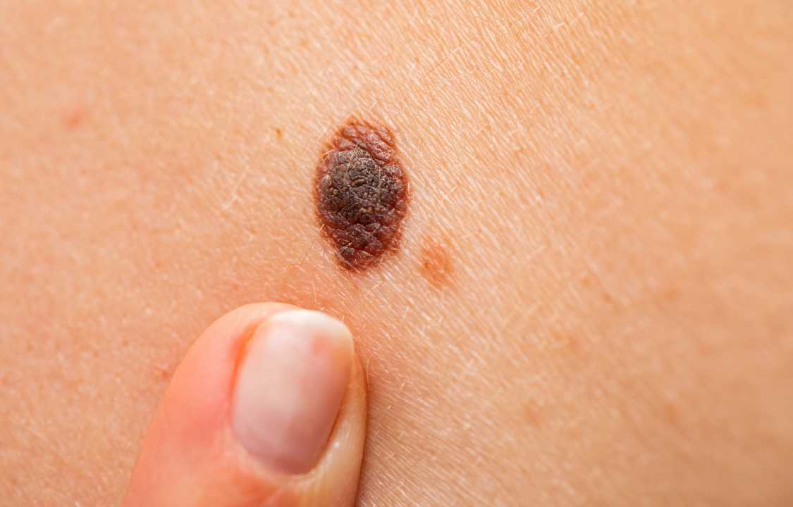
Stages of Melanoma, Explained by a Dermatologist
Oct
Survival rates of this serious skin cancer depend on which melanoma stage you have.
In the year 2022, the American Cancer Society (ACS) projected that over 96,000 individuals in the United States would receive a melanoma diagnosis. Melanoma stands as the most fatal type of skin cancer, but it’s important to realize that such a diagnosis does not equate to a death sentence. The prognosis and chances of survival hinge greatly on when melanoma is diagnosed and how promptly treatment commences.
Upon discovering melanoma, healthcare providers scrutinize the affected tissue under a microscope to gauge the extent of cancer in the body. They assess whether malignant cells have infiltrated other tissues or organs aside from the skin, a crucial step in determining the stage of the disease. In this comprehensive exploration, we’ll delve into the various stages of melanoma, their implications for treatment, and what they signify for prognosis.
Stage 0: Melanoma in Situ
The earliest stage of melanoma, labeled as stage 0 or melanoma in situ, is a Latin term that translates to “in position.” At this stage, cancerous cells exclusively inhabit the epidermis, which is the body’s outermost skin layer. The key characteristic of stage 0 melanoma is the absence of cancerous cell spread beyond the epidermis.
Treatment: The standard treatment for stage 0 melanoma involves wide excision surgery. During this procedure, healthcare providers surgically remove the affected skin, frequently a mole, and subsequently suture and dress the wound. This process typically includes excising a margin of normal skin around the affected area to ensure complete removal of cancer cells. Following surgery, the excised skin undergoes microscopic examination to ascertain the absence of any remaining cancerous cells.
In the case of stage 0 melanoma, where cancerous cells have not ventured into other tissues or organs, further treatment is typically unnecessary. In some instances, healthcare providers may consider alternative treatments such as radiation therapy, Mohs surgery, or the application of Imiquimod cream. However, it’s important to note that not all cancer experts unanimously endorse these treatments, with wide excision remaining the prevailing standard.
Survival Rate: Stage 0 melanoma boasts a favorable prognosis, with individuals who promptly receive treatment for localized melanomas (comprising stages 0, 1, and 2) exhibiting a five-year survival rate of 97%. In essence, they are approximately 98.4% as likely to survive five years post-diagnosis as individuals without these types of cancers.
Stage 1: Advancement Beyond the Epidermis
When melanoma progresses beyond the epidermis, it warrants a diagnosis of stage 1. In this stage, the tumor’s thickness does not exceed two millimeters, and it may exhibit ulceration, signifying penetration through the skin. Nevertheless, cancerous cells have not yet infiltrated nearby lymph nodes or organs.
Treatment: Treatment for stage 1 melanoma closely resembles that of stage 0. Healthcare providers surgically excise the affected area, suturing and dressing the wound. Following surgery, microscopic examination of the tumor ensures the complete removal of cancerous cells. Additionally, healthcare providers may opt for a biopsy of nearby lymph nodes to confirm the absence of melanoma spread.
Survival Rate: Stage 1 melanoma, when detected early and treated promptly, carries a five-year survival rate of 98.4%, akin to that of stage 0.
Stage 2: Moderate Tumor Thickness
In stage 2 melanoma, the tumor’s thickness ranges from one to four millimeters, potentially accompanied by ulceration as observed in previous stages. Just like in stages 0 and 1, cancerous cells remain confined to the primary tumor, with no signs of dissemination to nearby lymph nodes or organs.
Treatment: The standard treatment for stage 2 melanoma typically involves wide excision surgery. During this procedure, healthcare providers not only remove the melanoma but also excise a margin of surrounding skin, the size of which depends on the thickness and location of the tumor. Microscopic examination of the removed tissue confirms the absence of residual cancer cells. A biopsy of nearby lymph nodes may also be recommended in certain cases.
Survival Rate: Stage 2 melanoma, like its predecessors, falls within the category of localized melanomas. Consequently, individuals diagnosed with stage 2 melanoma boast a five-year survival rate of 98.4%.
Stage 3: Regional Spread of Cancerous Cells
Stage 3 melanoma marks the point at which cancerous cells infiltrate nearby lymph nodes or organs. Although the cancer extends beyond the primary tumor, there remains no evidence of metastasis to distant organs.
Treatment: Treating stage 3 melanoma typically necessitates more extensive measures than earlier stages. Healthcare providers may be required to remove not only the primary tumor but also enlarged lymph nodes upon palpation. This process involves a more substantial excision and frequently leaves a notable scar. A sample of the affected lymph nodes undergoes microscopic examination, with the removal of lymph nodes in the vicinity if melanoma cells are detected. Additional treatments may be recommended based on the healthcare provider’s findings, including surgery, radiation, immunotherapy, or participation in clinical trials.
Survival Rate: Stage 3 melanoma, encompassing regional spread but no distant metastasis, bears a five-year survival rate of 63.6%.
Stage 4: Distant Metastatic Melanoma
Stage 4 signifies the most advanced form of melanoma, characterized by the spread of cancerous cells to distant lymph nodes or organs such as the lungs, brain, or liver, a condition known as metastatic melanoma.
Treatment: The treatment approach for stage 4 melanoma aims at managing the disease, alleviating symptoms, and improving the patient’s quality of life rather than achieving a cure. Treatment strategies encompass surgical removal of the primary tumor and affected lymph nodes, radiation therapy, immunotherapy, targeted therapy, drugs used in clinical trials, and chemotherapy. While surgery may shrink tumors or alleviate symptoms, it is generally not curative. The emphasis is on controlling the disease and symptom relief.
Survival Rate: Stage 4 melanoma presents a formidable challenge for treatment, resulting in a five-year survival rate of approximately 22.5%.
Preventive Measures and Early Detection
Prevention and early detection are crucial in managing melanoma effectively. Understanding risk factors, such as exposure to ultraviolet (UV) light, fair skin prone to sunburns, a history of multiple blistering sunburns, numerous moles, atypical moles, or a family history of melanoma, empowers individuals to take proactive measures. Avoiding tanning beds and regularly applying broad-spectrum, water-resistant sunscreen with an SPF of at least 30 is recommended by the American Academy of Dermatology Association.
Monitoring the skin for abnormal growths and changes is essential for everyone, not solely those predisposed to skin cancer. Annual skin checks with a dermatologist or healthcare provider are integral components of proactive healthcare.
Conclusion
In conclusion, melanoma is a formidable form of skin cancer categorized into various stages, each carrying distinct implications for treatment and prognosis. The key to improving melanoma prognosis lies in early diagnosis and prompt treatment. Therefore, any alterations in moles or skin should not be overlooked, and immediate consultation with a healthcare provider is paramount. Melanoma’s aggressive nature underlines the importance of swift action when encountering suspicious skin changes. Furthermore, understanding risk factors and adopting preventive measures is pivotal in reducing the incidence of melanoma and safeguarding overall health. Regular skin checks are not mere precautionary measures but potentially life-saving practices, underscoring their significance in healthcare routines.