
Cardiovascular
Flurpiridaz will have a major impact on cardiac PET and nuclear imaging
Oct
For hospitals that have been on the fence about investing in PET imaging systems, the expected FDA clearance of flurpiridaz may be the missing piece of the puzzle that pushes many to make the investment. Bateman said facilities still need to plan ahead to line up funding and begin planning for an install.
“Those of us who have done this for years have learned that that’s a one- to two-year type business proposition. One of our initiatives through ASNC and Society of Nuclear Medicine and Molecular Imaging (SNMMI) is to begin encouraging people to understand what this agent is all about,” Bateman explained.
Why flurpiridaz is a game-changer in cardiac imaging
There are several key advantages that set flurpiridaz apart from existing radiopharmaceuticals. First, it has a longer half-life compared to currently available agents. This prolonged half-life facilitates unit dosing and exercise stress testing, offering clinicians greater flexibility and precision in diagnostic procedures. This alone represents a significant advancement in PET imaging.
“That’s a tremendous advance for the expansion of PET into the marketplace. The problem is that the penetration of PET into the broader community has been limited because you must have a hefty volume of patients on a daily basis to mitigate the risk of signing a contract for a rubidium generator that is very costly on a monthly basis,” Bateman said.
He said there are lots of studies and meta-analyses showing that PET is better than SPECT. It has better diagnostic accuracy, sensitivity and specificity, for example, as well as being associated with better image quality, more definitive interpretations and more confident interpretations. It is also much more accurate in particular subpopulations, like obese patients and women, where its has been shown to be superior over SPECT.
Being able to now order a unit dose from a commercial cyclotron and have it delivered is a major business game-changer.
“If you’ve got some patients, you order the tracer. So the risk has been switched, if you want to put it that way, from the provider to the manufacturer. This is an exciting time for people like me who have been practicing nuclear cardiology for quite some time. We haven’t had any new tracers since the early 1990s. That’s a long time ago. And along comes this flurpiridaz that has some very, very excellent characteristics. Not only is it a PET agent, but it’s also got a high extraction rate, particularly at high flows, which should substantially improve sensitivity. It’s a fluorinated F18 tracer. All of our PET cameras are designed specifically to be able to elicit excellent F18 images,” Batman said.
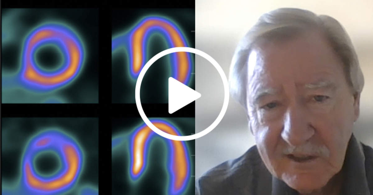
Flurpiridaz’s high extraction rate enhances sensitivity in detecting CAD. This characteristic ensures that even patients with more advanced or subtle forms of the disease can be accurately diagnosed. The tracer’s uptake by mitochondria in cells and its prolonged retention ensures that it does not redistribute, a critical feature for reliable and reproducible imaging, Bateman added.
One of the most remarkable aspects of flurpiridaz is its extended half-life of approximately 110 minutes. This attribute allows for stress testing with either pharmacological agents or exercise, offering clinicians more options in tailoring diagnostic procedures to individual patient needs. With traditional agents like rubidium, which have very short half-lives (about 75 seconds), exercise stress testing is often not feasible due to the limited time available for imaging. Flurpiridaz’s extended half-life mitigates this limitation, providing healthcare providers with a versatile tool for cardiac stress testing.
Flurpiridaz’s longer imaging window not only improves the overall image quality but also offers the opportunity to detect CAD at earlier stages, Bateman said. This is particularly significant as it allows for the identification of spatially relative perfusion defects, even before they reach the severity of traditional ischemic defects. In clinical practice, this means that physicians can potentially diagnose CAD in patients who may not exhibit severe symptoms or clear-cut perfusion defects, a capability that can significantly impact patient outcomes.